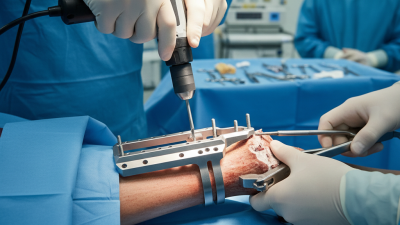
How to Choose the Right Proximal Humeral Plate

Choosing the right Proximal Humeral Plate begins with the patient, not the product catalogue. Fracture pattern, bone quality, tuberosity position, soft-tissue condition, and shoulder demands must guide the decision. A plate that fits a radiograph may still fail in a small, osteoporotic shoulder.

Orthopedic trauma expert Dr. Peter Boileau has emphasized a practical principle: “Treat the fracture pattern, not the X-ray alone.” This idea remains valuable when comparing plate length, screw trajectory, calcar support, locking options, and contour. The best design should restore alignment while respecting blood supply and rotator-cuff attachments. It should also allow controlled fixation of the greater and lesser tuberosities.

Experience matters here. Surgeons often assess the patient in multiple planes, review computed tomography, and compare the planned plate with the individual’s anatomy. A low-profile plate may reduce irritation, yet insufficient medial support can invite varus collapse. More screws do not automatically create better fixation. Placement matters.

Small details matter.

This article examines how to match a Proximal Humeral Plate to fracture morphology, bone strength, and surgical goals. It also considers practical concerns, including screw length, calcar purchase, plate position, and postoperative imaging. No implant is universally ideal. That is an important limitation. Surgeon training, patient expectations, and intraoperative findings can change the final choice. Readers should use these principles as educational guidance, not as a substitute for individualized evaluation by a qualified orthopedic specialist.
How to Choose the Right Proximal Humeral Plate

Understanding Proximal Humeral Fracture Patterns

How to Choose the Right Proximal Humeral Plate

Understanding Proximal Humeral Fracture Patterns

Plate selection starts with the fracture, not the implant tray. Obtain good radiographs and consider CT when fragments overlap or displacement remains unclear. Identify the surgical neck, anatomical neck, greater tuberosity, lesser tuberosity, and humeral head involvement. Pattern recognition matters.

A varus fracture may show medial column failure and poor calcar support. A valgus-impacted fracture can preserve some contact but still hide unstable tuberosities. Head-split fractures and fracture-dislocations demand careful assessment of articular damage and soft-tissue risk. Bone quality also changes the plan. Osteoporotic metaphyseal bone may require locking fixation, calcar screws, and sufficient plate length. Short plates can concentrate stress near the fracture.

Tuberosity position deserves close attention. The greater tuberosity influences cuff mechanics, while the lesser tuberosity affects subscapularis function. A plate should support these fragments without blocking their reduction. In practice, plate contour, screw direction, medial support, and the surgeon’s exposure must work together. A technically strong construct can still fail if reduction is poor.

One detail is easy to underestimate. The medial hinge may be thin or absent. Without restoring that support, screw fixation alone may not control varus collapse. I have learned that a familiar plate is not always the best choice. Rechecking fracture lines after reduction can change the decision. Sometimes the initial plan was simply too confident.

How to Choose the Right Proximal Humeral Plate

Understanding proximal humeral fracture patterns

Surgical-neck fractures are the most frequently encountered proximal humeral fracture pattern. Greater-tuberosity involvement, fracture displacement, medial cortical support, and head-split or articular extension should guide plate length, screw configuration, and the need for additional fixation. The percentages shown are approximate educational proportions synthesized from commonly reported clinical fracture-pattern distributions; actual frequencies vary by study population and classification method.

Assessing Patient Factors and Bone Quality

How to Choose the Right Proximal Humeral Plate: Assessing Patient Factors and Bone Quality

Choosing a proximal humeral plate begins with the patient, not the implant tray. Age, activity level, fracture pattern, and medical history all influence fixation demands. A younger patient may generate higher shoulder loads during recovery. An older patient may have fragile bone and limited healing potential. Diabetes, smoking, steroid use, and previous shoulder surgery also deserve careful review. These factors can change screw strategy, reduction goals, and follow-up planning.

Bone quality is often the difficult variable. Radiographs may suggest osteopenia, but they do not show every weak zone. Computed tomography can reveal thin cortices, comminution, and poor head support. Intraoperative assessment still matters. The surgeon should evaluate purchase, tuberosity integrity, and the risk of varus collapse. Plate length, screw direction, and support around the calcar should match the fracture and bone condition. More metal is not automatically better. Excessive hardware can irritate soft tissue or restrict movement.

Tips: Compare both shoulders when imaging is unclear. Check medial support before final fixation. Use multiple points of fixation in weak bone, while avoiding joint penetration. Record why the selected plate fits the patient’s anatomy. That reasoning improves communication and later review. A useful plan can still fail if reduction is poor. I have found that patient-specific judgment remains more reliable than a rigid checklist.

Comparing Plate Designs and Fixation Features

How to Choose the Right Proximal Humeral Plate

Plate design should match the fracture pattern, bone quality, and surgical approach. A narrow plate may reduce soft-tissue irritation, but it can offer less control in complex fractures. A broader plate may improve support, yet its profile can affect shoulder movement. Low-profile contours often help around the greater tuberosity. Still, positioning matters more than appearance. Even a well-designed plate can fail after poor reduction or weak medial support.

Fixation features deserve close comparison. Locking holes can improve screw stability in osteoporotic bone. Variable-angle holes allow more precise targeting around the humeral head. Inferomedial or calcar screws may support the medial column and reduce varus collapse. Some designs include suture holes for tuberosity repair. These details are useful, not magical. Screw length, trajectory, and purchase must be checked with imaging and surgical judgment. No plate design fits every fracture.

Tips: Compare the plate against preoperative images and the patient’s anatomy. Check whether screws avoid the joint surface. Confirm medial support before final tightening. Consider the incision and soft-tissue coverage. In my experience, small positioning errors create large mechanical problems. That lesson is easy to underestimate. Surgeons should also review fixation under multiple views, because one projection may hide a proud screw. A second assessment can change the plan.

How to Choose the Right Proximal Humeral Plate - Comparing Plate Designs and Fixation Features

Plate Design or Feature Typical Indications Fixation Options Angular Stability Key Advantages Important Limitations Selection Considerations
Anatomically Contoured Locking Plate Displaced or unstable proximal humeral fractures, including many three-part and four-part patterns. Multiple fixed-angle locking screws in the humeral head, with shaft holes that may accept locking or non-locking screws. High
Fixed-angle screws form a stable plate-screw construct.
Supports the humeral head in osteoporotic bone; precontoured shape can reduce the need for extensive plate bending; multiple screw trajectories can improve fragment control. A poorly positioned plate may cause subacromial impingement or screw penetration into the joint. Fixed trajectories may not suit every fracture line. Confirm plate height, tuberosity coverage, screw length, and the quality of medial column support before final fixation.
Variable-Angle Locking Plate Fractures requiring customized screw direction because of irregular fracture lines, small fragments, or limited safe corridors. Locking screws can be inserted within a manufacturer-specified angular range rather than only along one fixed trajectory. High
Angular stability is maintained when the approved range and insertion technique are followed.
Allows the surgeon to target dense bone, avoid fracture gaps, and adjust screw paths around the joint surface or other implants. The construct depends on correct technique, compatible screws, and adherence to the permitted angulation. Excessive off-axis insertion can reduce fixation reliability. Useful when standard fixed trajectories do not provide adequate purchase, but screw position must still be verified carefully with imaging.
Hybrid Locking and Compression Plate Fractures where both angular stability and controlled shaft-to-plate compression or adjustment are desirable. Combination of locking holes and conventional dynamic-compression or standard screw holes. High in locking holes
Conventional holes provide non-locking fixation where appropriate.
Offers flexibility: locking fixation can support weak metaphyseal bone, while non-locking screws can help draw the plate to the shaft or create compression in selected fracture patterns. Incorrect use of conventional screws may compromise reduction or create excessive compression. The plate should not be treated as a substitute for restoring medial support. Select when the fracture requires different fixation behaviors in the proximal segment and the shaft.
Low-Profile Plate Patients in whom soft-tissue irritation and prominence over the greater tuberosity or subacromial region are major concerns. Usually uses multiple proximal locking screws, with locking or standard shaft fixation depending on the design. High when locking holes are used Reduced plate prominence may decrease soft-tissue irritation and may be helpful when the deltoid and rotator cuff envelope is thin. A lower profile does not correct malposition. Limited plate surface or reduced screw spread may be less suitable for highly comminuted fractures. Prioritize accurate positioning below the greater tuberosity and assess whether the available proximal screw distribution is sufficient.
Extended-Length or Shaft-Extension Plate Fractures with distal extension, poor proximal shaft bone, long oblique patterns, or a need to span a weakened segment. Multiple shaft holes permit a longer working length and distributed fixation; proximal holes provide head and tuberosity fixation. High proximally
Shaft stability depends on plate length, screw distribution, and construct working length.
Spreads load over a larger area and can reduce stress concentration at the fracture site or near the end of the plate. May require a larger incision or more extensive soft-tissue handling. Excessive stiffness or inadequate working length can affect fracture healing mechanics. Choose a length that spans the involved region while avoiding unnecessary soft-tissue disruption and stress risers.
Calcar-Supporting Design Medial comminution, varus-collapse risk, osteoporotic bone, or fractures lacking reliable inferomedial cortical support. Inferomedial or calcar-directed screws, often combined with multiple humeral-head locking screws. High for medial support
Effectiveness depends on accurate screw placement and preserved bone.
Helps resist varus collapse and supports the inferomedial portion of the humeral head when the medial column is compromised. Calcar screws that are too short may not engage meaningful bone; screws that are too long may risk joint penetration. Consider this feature a priority in medial-column deficiency, especially when bone quality is poor or the head is tending toward varus.
Tuberosity Suture-Fixation Features Greater or lesser tuberosity fractures, rotator cuff attachment disruption, and fracture patterns requiring tuberosity reduction. Dedicated suture holes, eyelets, or slots for high-strength sutures placed through the rotator cuff tendon-bone junction. Supplementary
Sutures enhance fragment control but do not replace structural screw fixation.
Helps maintain tuberosity position, supports soft-tissue repair, and can distribute tensile forces from the rotator cuff. Suture fixation alone may not control a mechanically unstable fragment. Poor suture placement can cut through tendon or bone. Select a design with accessible holes positioned to permit secure fixation of both tuberosities without excessive tendon abrasion.
Non-Locking Compression Plate Selected fracture patterns with good bone quality, reconstructible cortical contact, and a need for conventional compression. Standard screws generate fixation through plate-to-bone compression and may provide dynamic compression in designated holes. Variable
Stability depends strongly on bone quality, screw purchase, and cortical contact.
Can provide direct plate-to-bone fixation and controlled compression when the fracture pattern and bone stock are suitable. Less effective in osteoporotic metaphyseal bone and less capable of maintaining a fixed-angle relationship between the screws and the humeral head. Use selectively; it is generally less forgiving when there is metaphyseal comminution, poor bone quality, or limited cortical support.
Screw-Length and Joint-Safety Features All proximal humeral plating procedures, particularly fractures with a collapsed or irregular humeral head. Multiple screw lengths, depth-control options, radiographic markers, and guided insertion systems may be available depending on the plate design. Essential safety feature Helps maximize purchase while reducing the risk of intra-articular screw penetration, a recognized complication of proximal humeral fixation. Even with guides and markers, fracture reduction can change after fixation; intraoperative imaging and final screw-length assessment remain necessary. Prefer systems that support accurate depth measurement and provide clear imaging of the humeral head in multiple views.

General comparison only. Final plate selection should be based on fracture morphology, bone quality, medial-column integrity, soft-tissue condition, imaging, and the fixation technique appropriate for the individual patient.

Selecting Plate Position and Surgical Approach

How to Choose the Right Proximal Humeral Plate

Selecting plate position and surgical approach begins with the fracture pattern, not the implant outline. Review true anteroposterior, scapular Y, and axillary images, adding CT when fragments overlap. Note the medial column, tuberosity displacement, head-split features, and bone quality. These details guide both exposure and fixation strategy.

A lateral plate usually sits slightly posterior to the bicipital groove and below the greater tuberosity. Excessive height can cause painful subacromial contact during abduction. Excessive posterior placement may weaken control of the tuberosity. The deltopectoral approach offers familiar access to the anterior shoulder and bicipital groove. An anterolateral approach can provide direct lateral exposure, but the axillary nerve demands careful protection and measured dissection. Keep the incision and retraction respectful of soft tissues.

Intraoperative fluoroscopy should confirm plate height, screw length, head position, and joint clearance. A guidewire that appears safe in one view may enter the joint in another. That mistake is preventable, yet it still occurs when imaging is rushed. Patient position also matters; beach-chair setup can improve visualization, while arm manipulation must remain controlled. An experienced surgeon should adapt the approach to fracture biology, skin condition, and training. A templated plan is useful, but not sacred. Sometimes the safest decision is to change exposure after the first fragment is inspected.

Confirming Compatibility, Stability, and Surgical Readiness

How to Choose the Right Proximal Humeral Plate

Confirming Compatibility, Stability, and Surgical Readiness

Choosing the right proximal humeral plate starts with compatibility, not appearance. Confirm that it matches the fracture pattern, humeral anatomy, and fixation plan. Review radiographs and, when needed, CT images before opening the set. Measure the proximal humerus carefully. Small differences matter.

Check screw trajectories against the humeral head, calcar, and medial cortex. Ensure each hole supports the intended locking or compression strategy. Bone quality also changes the plan. Osteoporotic bone may require more fixation points and careful torque control.

Stability depends on more than plate strength. Position the plate below the greater tuberosity and away from the rotator cuff. Confirm that screws will not enter the joint. Use depth measurement and live imaging when appropriate. A trial plate can reveal problems early. In practice, I have seen a plate fit the model but fail to sit flush during surgery. Anatomy is rarely perfect. Recheck reduction after provisional fixation, because a stable-looking construct can still hide varus collapse risk.

Surgical readiness includes small details. Verify the instrument set, drill guides, screw lengths, backup options, and imaging access. Inspect threads, guides, and sterile packaging before incision. Do not rely on memory. A missing guide can interrupt a carefully planned procedure. Record the selected plate size and planned screw positions, while allowing changes when findings differ. The best choice may change after exposure. That requires disciplined reassessment.